Monday, 3 May 2010

Analyse the relationship between smoking and coronary heart disease and lung cancer.

Cardiovascular diseases affect the heart and arteries causing ill health and is also the leading cause of death in the u.k. An estimated 198,000 people are dying every year in the u.k of (CVD). (CVD) links to smoking include:- High blood pressure, Coronary heart disease, Angina, Atherosclerosis, Heart failure and Heart attacks.


Cerebrovascular diseases affect the flow of blood to the brain and is the third largest cause of death in the u.k , they include 'Dementia' and 'strokes'. In England 111,000 people have strokes every year.

Smoking is thought to be the cause of 85-90% of lung cancer and is the leading cause of cancer related deaths. In 2007, there were 29,660 deaths in England and Wales.

Social smoking ( only smoking on occasions) still increases the risk of coronary heart disease and cancer-30% more than none smokers.

Research has shown that smoking 4 joints (tobacco mixed with marijuana) may cause as much damage as 20 cigarettes.

Second hand or passive smoke contains 4,000 chemicals and of those 69 of them are carcinogenic. Passive smoking carries the increased risk of lung cancer (24%) and heart disease(25%). It also gives a higher chance of blood clots due to the blood becoming sticky.

Ref:-Image from www.scandirectory.com/blog/smoking_Heartdisease_Scan_Blog_June09.jpg&imgrefur

www.nhs.uk/conditions/smoking-(quitting)/pages/risks.aspx ( last reviewed 13/04/10) (accessed on 03/05/2010)

www.nsh.uk/chq/pages/2289.aspx?categoryid=53&subcategoryid=536 (last reviewed 29/11/2007) (accessed 03/05/2010)

Discuss the relationship between diet, blood pressure, blood cholesterol and circulatory disease.


Too much fat in the diet can cause fatty build ups of plaque in the arteries called Atheroma. The more blocked an artery is the less space there is for blood to flow through it , resulting in higher blood pressure, as the heart has to work harder to circulate the blood and at the same time highers the stress on itself.

An important part of reducing cholesterol in the blood is eating a healthy diet, this includes eating less saturated fats and trans fats, eating more fruit and veg, whole grains and fibres,drinking less alcohol and reducing salt intake.

Trans fats naturally occur in meat and dairy products. They can be found in many processed foods and sweets and have the same effects on the body as saturated fats.

However, there are fats that the heart needs to stay healthy. These are mono and poly-unsaturated fats which are found in fish oils and plants. By lowering triglycerides in the blood, they lower the risk of cardio vascular disease (CVD) by reducing blood clotting and regulating the heart rhythm.

Some plant derived compounds-'stanol' or 'sterol esters' have been shown to reduce cholesterol levels. They can be found in some drinks, yoghurt's, spreads and soya(dairy alternative).

A person should try to consume 5 portions of fruit and veg a day. They contain essential nutrients eg: vitamins C and E and carotenoids.(organic pigments). They could help protect the body from (CVD) by limiting the damage done to the blood vessels by cholesterol.

Research has shown that wholegrain foods can reduce the risk of (CVD) by up to 30% . While Fibre lowers LDL cholesterol.

High intakes of alcohol results in higher risk of clots forming in the blood. However 1-2 units of alcohol a day has been found to reduce the risk of (CVT). Saving all the units up and then binge drinking does not give the same benefits.

Soya protein is a good substitute for meat and 25g+ a day has shown a reduction in LDL cholesterol and (CVD). It inhibits the growth of cells that form artery clogging plaque.

Ref: Image from www.kidneydiettips.dowitablogs.com/up-content/uploads/2008/10/istock_000006148437-heart.jpg

www.bbc.co.uk/health/treatments/healthy-living/nutrition/dietary_cvd.shtml
(reviewed in July 2008 by MRC Human Nutrition Research. (accessed 03/05/2010)

Sunday, 2 May 2010

Describe the changes in artery structure associated with circulatory disease.


Atheroma (plaque) is the main cause of cardiovascular disease such as heart attacks, strokes and angina.It is also referred to as atherosclerosis and hardening of the arteries.

It is a build up of fatty deposits inside the lumen of the artery. If left untreated Atheroma can build up over time and partially or fully block arteries resulting in ill health and death.

Sometimes a minute crack on the inside wall of the blood vessel can cause a blood clot to form over the Atheroma, and this may block the artery completely.

Ref:Image from www.texasheat.org/HIC/Topics/Cond/pud.Cfm

www.patient.co.uk/health/Atheroma.htm (Last reviewed 15/04/2010) (accessed 02/05/2010)

Eplain the process for redistributing blood during exercise.


When the body is at rest aprox 15% of blood is supplied to the muscles and the rest goes to the organs.
When the body starts exercising this per cent age can increase to 80-85% and less blood goes to the organs all thoe the amount of blood that goes to the brain stays the same.

Blood being redistributed around the body is called the 'vascular shunt mechanism'

During light exercise redirection of blood to the skin enables the body to get rid of heat and keep cool. Skeletal muscle arterioles and pre-capillary sphincters vasodilate, increasing blood flow to muscles being used and organ arterioles and pre-capillary sphinchters vasoconstrict, temporally reducing blood flow to the organs.


Ref: Image from www.brainbasedbusiness.com/uploads/brainexercise-thumbjpg

www.webcache.googleusercontent.com/search?g=cache:12brQbSNO-UJ:www.school-portal.co.uk/GroupDownloadfile.asp%3FGroup (Accessed 02/05/2010)

Explain the mechanisms for regulating ventilation and pulse rates.


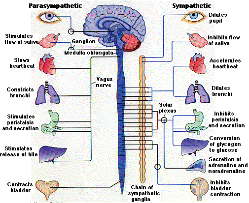
Ventilation and pulse rates are controlled by the nervous system . The nervous system consists of the brain and the spinal cord. Electrical impulses are transmitted around the body by neurones to enable the body to deal with certain situations.There are two sides to the nervous system known as the sympathetic nervous system(which speeds things up) and the parasympathetic nervous system(which slows things down).
This process can be seen during times of fear and stress, and is often referred to as 'Fight or Flight'.This means that when the brain senses danger, the sympathetic nervous system, will send electrical impulses down the spinal cord and along neurones which will : Dilate the pupils (for better vision) , inhibit saliva flow, Dilate the bronchi (to allow for maximum amount of oxygen to enter the body), inhibit peristalsis and secretion, Convert glycogen to glucose, secrete adrenaline (which increases heart rate and breathing) and inhibits bladder contraction.
When the danger has passed then the para-sympathetic nervous system takes over and all the reactions above return to normal.

Calculate Cardiac output and discuss the importance of this valve.


The' Cardiac output' (CO)is the amount of blood pumped per minute by the heart. In men this is aprox 5 lpm and in a woman it is aprox 4.5 lpm.
The' Pulse'(HR) is a way of measuring the number of cardiac cycles per minute. It normally ranges between 60-80 bpm.
The' Blood pressure' is the intensity of the blood when it leaves the heart eg: 120/80.
The 'Stroke volume' is the amount of blood per heart beat.
Measurement of the cardiac out-put is important as a change in the (CO) may indicate a change in health. Each (CO) would be assessed on the individual and their circumstances.
Access to HE class tutorial.

Explain the electrical activity of the heart during a heart beat.

The heart is stimulated by the 'Sympathetic' and 'Para-sympathetic' nervous system. The Sympathetic nervous system speeds the heart up while the Para-sympathetic nervous system slows it down.
The heart has its own electrical pace maker that is in the upper part of the Right atrium(RA). It is a collection of electrical fibres that are highly specialised called the 'Sino-Atrial (SA) node.

The (SA) node creates a number of sparks per minute but this can be increased during exercise or illness and lowered during rest and when a person is very fit.

The (SA) node fires an electrical impulse which causes the Right atrium to contract. This is called A 'P' wave on an electrocardiogram (ECG). The electrical impulses then move to the ' Atrio-ventricular' node (AV) node where it pauses very briefly to allow blood to finish emptying from the Atrium into the Ventricle. This pause is called the 'PR interval' on a ECG. Next the eletrical impulses travel through the Left and Right Bundle Branches, also know as 'His fibres' and 'Purkinje fibres'. This makes them contract and blood is pumped into the Pulmonary artery and the Aorta. On an electrocardiogram this is called the 'QRS complex'. As the Ventricles recover it produces a 'ST segment' and a 'T wave' on the ECG. Ref: Image from@www.sites.google.com/site/ehgprojectgmu/

Dr. Abdulla.M @ www.heartsite.com/html/eletrical_activity.html. Last reviewed 04/04/2010. Accessed 14/04/2010.